

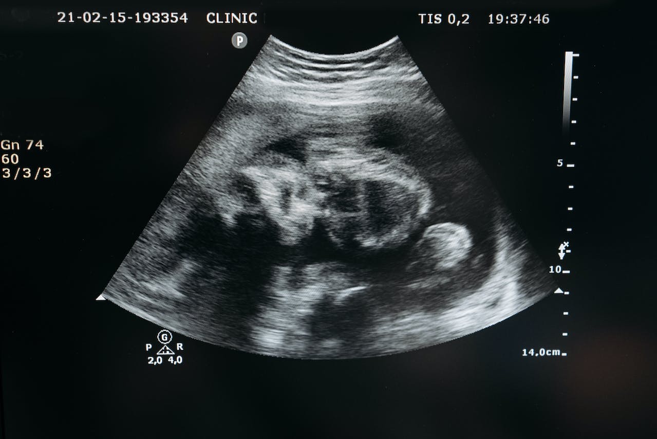
Ultrasound Scan In Pregnancy
Introduction
Thank you for reading this post, don't forget to subscribe!An ultrasound machine is a machine that uses extremely high sound waves inside our bodies to create images on a monitor. An ultrasound scanning is a procedure done to look for problems inside our bodies. In a pregnant woman, however, the scan is done to look for conditions of a pregnancy in both the normal and abnormal situations, at any given point in the duration of the pregnancy.
In normal pregnancy situations, ultrasound scans can help us determine
- Gestational age (age of the pregnancy)
- Baby size (can also determine if the baby is growing normally)
- Tentative delivery date
- Number of babies (one or more)
- The gender of the baby
- Lie (how the fetus is lying)
In abnormal situations, ultrasound scans can help us diagnose
- Position (whether inside the uterus or not – ectopic pregnancy)
- Type of pregnancy (whether normal or a mole)
- Miscarriages
- Displaced or low-lying placenta (Placenta Previa)
- Congenital abnormalities like Down’s Syndrome
What to expect
Every pregnant woman is ideally expected to have at least two ultrasound scans in an index pregnancy. The first is done as early as possible around the time the woman missed 2 consecutive periods, which is about two months into the pregnancy. This helps for an accurate determination of the expected delivery date (EDD), to rule out ectopic pregnancies and moles. The second one is done after the pregnancy has transited midway, say around the 5th month to see how the baby is doing and to diagnose any abnormality. It can also help check for the location of the placenta, how the baby is lying, i.e. whether normal or breech, and also the sex or gender of the baby.
The scan procedure is painless and does not require any special preparations, except that some sonographers advise having a full bladder in early scans. You lie down on your back on a couch and expose only your belly from the breastbone to the urinary bladder just above the pubic hairline.
The sonographer will apply a gel on your tummy which may feel cold, and then use a probe to go around the abdomen to capture all the images. You may politely request to view what he or she is viewing so that you can also appreciate what is being done. After the scan, a piece of tissue paper is provided to clean up the gel before you adjust your dress. The scan is printed and is either given to you or attached to your file records.
After the scan
In most cases, the scans are right, but in some cases, a problem among those listed above may be seen. If that is the case, the sonographer will notify you and discuss the appropriate line of management with you. For instance, if a mole is seen rather than a pregnancy or a pregnancy is seen outside of the uterus, they are evacuated or surgically removed respectively; if the baby is not lying straight, an offer to turn or rotate it is provided; if a congenital anomaly is seen, an option of termination or keeping on with the pregnancy is given. So make sure you ask if your scans are good after it is done.


